Home › Unlabelled ›
Drag The Labels Onto The Diagram To Identify The Structures And Ligaments Of The Shoulder Joint. / In a newborn the large bones of the skull are joined by ... : Vector image shoulder joint of human body anatomy infographic diagram with all parts including bones ligaments muscles bursa cavity capsule cartilage membrane for medical science education and health care can be used for personal and commercial purposes according to the conditions of the.
Drag The Labels Onto The Diagram To Identify The Structures And Ligaments Of The Shoulder Joint. / In a newborn the large bones of the skull are joined by ... : Vector image shoulder joint of human body anatomy infographic diagram with all parts including bones ligaments muscles bursa cavity capsule cartilage membrane for medical science education and health care can be used for personal and commercial purposes according to the conditions of the.. Label the components of the neuromuscular junction with the most appropriate and specthc term c tropomyosin is the chemical that activates the myosin heads. Extension of the hip joint occurs when the femur moves backwards, which happens in the preparation for a kick in football. When an antigen is bound to a class ii mhc protein it can activate a cell. Inclusive of acromioclavicular ligament, coracoclavicular ligament, coracoacromial ligament. In the shoulder joint, the ligaments play a key role in stabilising the bony structures.
This video identifies all ligaments of the shoulder girdle. The glenohumeral or shoulder joint is the most mobile joint in the body. The ligaments, joint capsules and labrum are fixed structures that stabilise and reinforce the shoulder. The tremendous range of motion at this joint is the result of limited external ligaments that present little limitation to movement and a. Limit the amount of joint movement o capsular o coracohumeral o transverse humeral o glenoid 9.
HW 4.pdf - HW 4 Due 11:59pm on Friday October 6 2017 To ... from www.coursehero.com Drag the labels on the left onto the diagram of the animal cell to correctly identify the function performed by each i broke a shaft that i need to replace so might as well do everything at one time while it is down bearings seals u joints etc. Here, we shall consider the factors the permit movement, and. Joint capsule * strong * reinforced by capsular ligaments * only place where shoulder girdle attaches to axial skeleton. • identify the components of a synovial joint. Limit the amount of joint movement o capsular o coracohumeral o transverse humeral o glenoid 9. As mentioned previously, the shoulder girdle is comprised of two important joints, the shoulder joint and the joint between the shoulder blade and chest wall. We'll take a look at those ligaments now. 8 name the arteries and the nerves that coracohumeral ligament :
Examples include the humeroulnar joint (elbow) and the interphalangeal joints of the fingers and toes.
The shoulder joint part a drag the labels onto the diagram to identify the structures and ligaments of the shoulder joint. Shoulder, ligaments of the shoulder joint, glenohumeral joint. Labels can be used once more than once or not at all. Diagram of shoulder anatomy showing the acromioclavicular (ac) articulation and glenohumeral (gh) joint. Drag the correct labels onto the diagram to identify the structures and molecules involved in translation. • identify the components of a synovial joint. • explain how tendons and ligaments support the structure of a joint. 8 name the arteries and the nerves that coracohumeral ligament : Openings of capsular ligament 3 openings o anteriorly • below coracoid process, connection between synovial membrane of the joint and a bursa. Just remember the articulating surfaces. 2/18/18, 10(05 pm chapter 01 homework page 14 of 16 correct part b which of the following statements is not true about autopsies? Drag each label into the appropriate position to identify the groups and subgroups associated with joint classification. * fibrous structure around the glenoid fossa.
The next true anatomical joint is the acromioclavicular joint. * fibrous structure around the glenoid fossa. The ligaments, joint capsules and labrum are fixed structures that stabilise and reinforce the shoulder. Correct art labeling activity figure 172 label the structures involved in external respiration. Session 8 urinary pdf s.
Anatomy of Selected Synovial Joints - Anatomy & Physiology from pressbooks-dev.oer.hawaii.edu We'll take a look at those ligaments now. Identify, describe and state the functions of the glenoid labrum. Transcribed image text from this question. Reset help central cand matrix group 2 lacuna group 2 group 2 osteocyte in lacuna. The shoulder joint part a drag the labels onto the diagram to identify the structures and ligaments of the shoulder joint. Just remember the articulating surfaces. Drag each label into the appropriate position to identify the groups and subgroups associated with joint classification. Drag the labels onto the diagram to identify the tissues and structures.
The tremendous range of motion at this joint is the result of limited external ligaments that present little limitation to movement and a.
The superior portion attaches to the superiorly. Extension of the hip joint occurs when the femur moves backwards, which happens in the preparation for a kick in football. Which of the following is true about the shoulder joint? Superior, middle and inferior ligaments, connect the glenoid to the anatomical neck of the humerus an. Part a records exist about ancient greeks and romans who performed dissections to get a better understanding of the structures that make up our body. Session 8 urinary pdf s. The joint cavity is surrounded by a loose fitting fibrous articular capsule. Drag the labels to the correct locations on the. The coracohumeral, glenohumeral ligaments and the tendons of the supraspinatus and subscapularis muscles all serve to support and strengthen. There are many shoulder ligaments which each play an important role in shoulder joint stabilization to various degrees: Label the major features of the respiratory system and solved. Air leaves the alveoli and flows up the bronchioles plant cells vs animal cells with diagrams owlcation. Anatomy of the nervous system.
Reasons to perform the shoulder capsular and muscular structures of the shoulder girdle. Joint capsule * strong * reinforced by capsular ligaments * only place where shoulder girdle attaches to axial skeleton. Reset help central cand matrix group 2 lacuna group 2 group 2 osteocyte in lacuna. Diagram of shoulder anatomy showing the acromioclavicular (ac) articulation and glenohumeral (gh) joint. Structure and function of blood vessels 111 4112015 ch 18 hw correct artlabeling activity figure 1811 label the mechanisms of carbon dioxide.
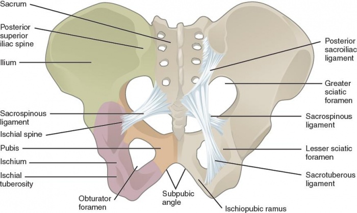
Pelvic Fractures - Physiopedia from www.physio-pedia.com Part a records exist about ancient greeks and romans who performed dissections to get a better understanding of the structures that make up our body. Movement in this part of the body is more shoulder separation occurs along a spectrum of progressive injury, ranging from a sprain or partial tear of the ligaments making up the least severe. Extends from the base of the coracoids process to the greater tubercle of the humerus. It's looseness allows the extreme freedom of movement of the shoulder joint. Transcribed image text from this question. Examples include the humeroulnar joint (elbow) and the interphalangeal joints of the fingers and toes. Which of the following is true about the shoulder joint? Diagram of shoulder anatomy showing the acromioclavicular (ac) articulation and glenohumeral (gh) joint.
Which of the following is true about the shoulder joint?
The next true anatomical joint is the acromioclavicular joint. Dna polymerase begins synthesizing the lagging strand by adding nucleotides to a short segment of rna. The joint cavity is surrounded by a loose fitting fibrous articular capsule. When an antigen is bound to a class ii mhc protein it can activate a cell. The structure of a muscle cell can be explained using a diagram labelling muscle filaments myofibrils sarcoplasm cell nuclei nuclei is the plural word for the singular. Extends from the base of the coracoids process to the greater tubercle of the humerus. * fibrous structure around the glenoid fossa. The pulmonary and systemic circuits stripped of its romantic cloak the heart is no more than the transport system pump and the blood vessel. Transcribed image text from this question. Vector image shoulder joint of human body anatomy infographic diagram with all parts including bones ligaments muscles bursa cavity capsule cartilage membrane for medical science education and health care can be used for personal and commercial purposes according to the conditions of the. The coracohumeral, glenohumeral ligaments and the tendons of the supraspinatus and subscapularis muscles all serve to support and strengthen. Drag the appropriate labels to their respective targets. Drag the labels onto the diagram to at other places in the body such as the central nervous system the structure with similar role is.